Chapter 11Scanning Electron Microscopy
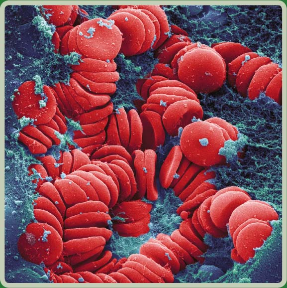
This photomicrograph features a colored scanning electron micrograph (SEM) revealing the clotting of red blood cells collected from an eighteen-year-old patient. The red blood cells were just beginning to clot when this image was made. The image magnification was x2990 when printed at a distance of 10 cm wide. Image courtesy of Ted Kinsman.
Introduction
The primary use of scanning electron microscopy (SEM) is to observe topographical structures leading to the production of images with large depth of fields at relatively high magnifications. Current machines are now capable of performing ...
Get Laboratory Imaging & Photography now with the O’Reilly learning platform.
O’Reilly members experience books, live events, courses curated by job role, and more from O’Reilly and nearly 200 top publishers.

