Chapter 10Confocal Microscopy
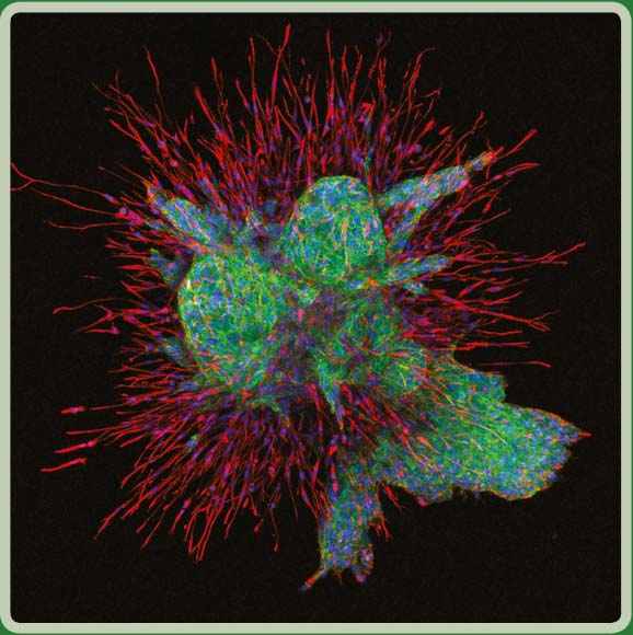
This photomicrograph features a human oral cancer cell line growing in the form of a spheroid. This culture technique avoids artificial adhesion of cells to a culture dish and promotes the cell—cell interactions that naturally occur among tumor cells. The spheroid is embedded in a matrix of type I collagen, which models the normal barriers encountered by oral cancer cells as they invade destructively into adjacent tissues. Immuno-fluorescent staining was used to show E-cadherin (alexafluor 488, green) and vimentin (alexafluor 568, red). Cell nuclei are imaged in blue (DAPI). The image was captured ...
Get Laboratory Imaging & Photography now with the O’Reilly learning platform.
O’Reilly members experience books, live events, courses curated by job role, and more from O’Reilly and nearly 200 top publishers.

