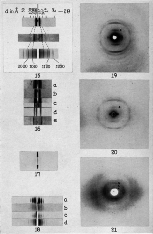
As early as 1885 Adolf Mayer showed that mosaic disease of the tobacco plant is contagious; we now know that it is caused by tobacco mosaic virus. Martinus Beijerinck (1851–1931) further isolated a “contagium vivum fluidum” (virus) from tobacco leaves, distinguishing the causative agent from bacteria. Due to their small size, almost all viruses cannot be visualized by conventional microscopy. Beginning in the 1930s Helmut Ruska pioneered the use of the electron microscope to visualize viruses (Kruger et al., 2000). Early studies of the structure of viruses based on x-ray crystallography were performed by John D. Bernal (1901–1971). He also trained Maurice Wilkins and Rosalind Franklin (who confirmed the structure of the double helix of DNA) and Nobel laureate Dorothy Crowfoot Hodgkin (who solved the structure of vitamin B12). Together with Rosalind Franklin, Bernel studied tobacco mosaic virus in the 1950s. Bernal and Fankuchen 1941 obtained a variety of purified viruses and performed x-ray analyses. This set of images shows figures 15 (demonstrating shifts of intermolecular reflections), 16 (showing varying concentrations of viruses), 17 (enation mosaic virus), 18 (dry gels of various virus proteins), 19 (tobacco mosaic virus), 20 (cucumber mosaic virus), and 21 (potato virus X).
14
Completed Genomes: Viruses
INTRODUCTION
In this chapter we will consider bioinformatic approaches ...
Get Bioinformatics and Functional Genomics, Second Edition now with the O’Reilly learning platform.
O’Reilly members experience books, live events, courses curated by job role, and more from O’Reilly and nearly 200 top publishers.

